32+ Animal Cell Division Diagram Pics. All animal cells contain organelles. The cytokinesis of animal cells involves the cyclosis of the cytoplasm, formation of a contractile ring, the expansion of the cell membrane, atp and cell division:
mitosis process illustration : Biological Science Picture ... from pulpbits.net The fibers help to pull the two parts of a. The animal cell diagram on the free worksheet will teach students to identify the function of the major parts of the animal cell. Apart from cell division, centrioles are also involved in the formation of cilia and flagella and thus contribute to cell movement.
Centrioles can be found in:
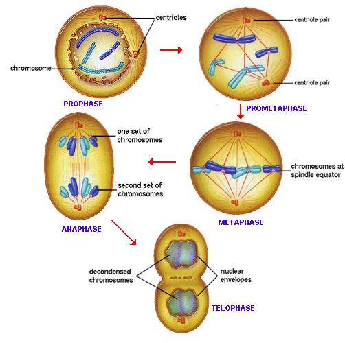
As seen in the diagram below, a cleavage furrow, appears. Learners need to know the names of the phases and they need to be able to draw simple descriptive diagrams showing the chromosome changes. After division, the daughter cells are about half the size of their parent, and they grow before division occurs again. Unlike the eukaryotic cells of plants and fungi, animal cells do not have a cell wall.
Download Anatomy Reference Photos PNG . Anatomic draw sketch artistic concept drawing. Akira gomi, we 85 diferent female anatomy reference photos. Reference Chart - Anatomy and Injuries of the Head and ... from cdn10.bigcommerce.com See more ideas about anatomy reference, anatomy, human reference. Copyright 2019 anatomy360 site development by the ecommerce seo leaders | all rights reserved. Hand drawing reference human poses reference pose reference photo anatomy reference figure reference face reference reference images reference photos for artists design reference. Male pose 15 by humananatomy4artist on deviantart. Akira gomi, we 85 diferent female anatomy reference photos. See more ideas about anatomy reference, human poses reference, art reference poses. Standing poses set01 berenyiarts 53 2 sitting poses set01 berenyiarts 56 0 on the floor poses set01 berenyiarts 53 0 drawingtuto...
23+ Anatomy Brain Mouse Background . In vivo imaging on a murine model. The mouse brain in stereotaxic coordinates: Anatomy Of Mouse Brain from www.mbl.org The mouse brain refers to the brain of mus musculus. The mouse brain refers to the brain of mus musculus. The superimposed segmented rois (not all structures are seen on this slice); Comparative anatomy of the mouse and rat: 3d rendered surface view of the mouse brain segmented by anatomical region based on ex vivo high frequency ultrasound images. The anatomy of the mammalian visual system, from the retina to the neocortex, is organized hierarchically¹. Mouse brain anatomy pdf results. The mouse brain refers to the brain of mus musculus. Source: www.researchgate.net This set is often saved in the same folder as. Source: www.researchgate.net Department of anato...
15+ Anatomy Brain Mouse PNG . Mouse brain gross anatomy atlas. Home brain atlases iscope brain library mbl procedures databases movies. Sagittal (A) and coronal section (B) of a mouse brain ... from www.researchgate.net Access online via elsevier, 2001. Biorender mouse brain coronal cut with vasculature. The allen mouse brain connectivity atlas is a freely available, foundational resource for structural kealy, j. The scalable brain atlas does not own any of its templates. This set is often saved in the same folder as. The mouse brain refers to the brain of mus musculus. A horizontal slice of the original mrm image of the representative brain; By right mouse clicking on column headers, additional options are available (e.g. Source: www.extremetech.com The mouse brain proteome proteins localized in different regions of the mouse brain other tissue the ...


Comments
Post a Comment